

Cartilage research often depends on detecting subtle changes in extracellular matrix composition before gross tissue structure is visibly altered. Safranin o staining is widely used in research to visualize cartilage proteoglycans, helping investigators compare matrix-rich regions with areas where glycosaminoglycan content has been reduced.
That information is valuable because matrix remodeling rarely involves only one component. Proteoglycan loss, collagen deposition, fiber reorganization, and structural damage can occur together, so choosing complementary stains can provide a more complete view of how tissue is changing.
Look Beyond General Morphology
Routine H&E staining gives useful information about nuclei, cellular organization, and overall architecture. However, it does not specifically show how much proteoglycan remains in cartilage or how collagen fibers are distributed within connective tissue.
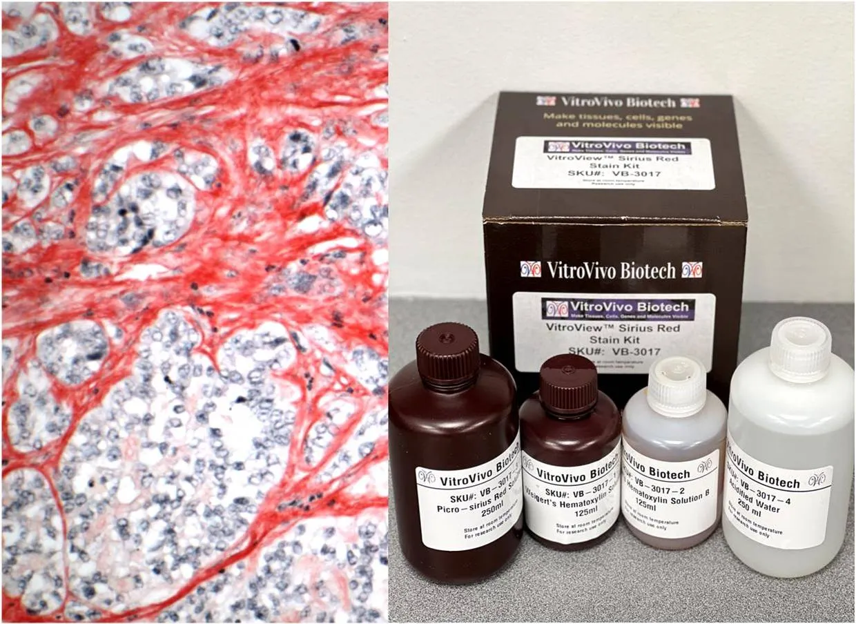
When collagen becomes a major part of the research question, a Picro Sirius Red stain can provide a clearer view of collagen I and III fibers. Under standard light microscopy, collagen appears red, while polarized light can reveal birefringence patterns that help distinguish thick and thin fiber populations.
Why Proteoglycan Loss Matters
Proteoglycans contribute strongly to the mechanical properties of cartilage by attracting water and supporting resistance to compression. Changes in their abundance can therefore signal matrix deterioration even when the tissue still appears relatively intact on a routine section.
Safranin O is a cationic dye that binds to polyanionic tissue components. In cartilage research, stronger orange-red staining generally reflects greater proteoglycan content, while reduced intensity may indicate depletion that should be interpreted alongside morphology and experimental context.
Collagen Adds Another Layer
Collagen provides structural strength across many tissues, including cartilage, liver, kidney, muscle, and fibrotic lesions. Measuring its distribution can help researchers assess remodeling, scar formation, or extracellular matrix expansion in experimental models.
Picro-Sirius Red is especially useful when collagen organization matters as well as presence. Polarized-light evaluation can add information about fiber thickness and orientation, making the stain valuable in studies where matrix architecture is changing over time.
Plan Adjacent Sections Carefully
Researchers can often gain more information by staining adjacent sections from the same block. One section may be used for routine morphology, another for proteoglycan assessment, and another for collagen-focused analysis.
This strategy conserves tissue while allowing related measurements to be compared within similar anatomical regions. Because adjacent sections are not perfectly identical, consistent orientation and careful region matching are still important when interpreting differences across slides.
Keep Technical Variables Stable
Section thickness, fixation, decalcification, deparaffinization, staining time, differentiation, and mounting can all affect final color intensity. Standardizing these steps is essential when researchers intend to compare staining between experimental groups.
For cartilage specimens, decalcification deserves particular attention because harsh conditions can alter tissue chemistry. Detailed records of processing conditions help investigators determine whether an unexpected staining change is biological or introduced during preparation.
Use Controls and Defined Scoring
A known control tissue can confirm that staining chemistry is performing as expected. Controls are especially useful in multi-batch studies, where reagent age, timing, or handling differences may otherwise complicate comparisons.
Researchers should also define scoring methods before reviewing the complete dataset. Options may include visual grades, positive-area measurements, optical-density analysis, or region-specific assessments, depending on the biological question and imaging workflow.
Standardize Image Acquisition
Microscope settings influence how stained tissue appears in recorded images. Magnification, illumination, exposure, white balance, and polarization settings should remain consistent when images will be compared across groups.
Consistent acquisition becomes even more important when digital measurements are planned. Changes introduced by the microscope or camera can affect apparent staining intensity, so imaging should be treated as a controlled part of the experiment rather than a final documentation step.
Interpret Matrix Features Together
Proteoglycan depletion and collagen remodeling should not automatically be treated as equivalent events. One may change earlier or more strongly than the other, depending on tissue type, model, treatment, and stage of the biological process.
The most useful interpretation comes from comparing multiple matrix features with tissue morphology and experimental design. Combining complementary stains helps researchers build a richer picture of extracellular matrix change without expecting one method to answer every structural question.
Conclusion
Extracellular matrix research benefits from methods that reveal specific tissue components rather than only overall morphology. Proteoglycan-focused and collagen-focused stains provide different information, allowing investigators to examine matrix composition and structural organization from complementary perspectives.
Careful section planning, standardized processing, appropriate controls, and consistent imaging strengthen those comparisons. When staining methods are matched to clear research questions, valuable cartilage and connective-tissue specimens can provide more informative and reproducible data.